Vital Signs
Vital Signs. Nursing 125. Vital Signs. Temperature, pulse, respiration, blood pressure (B/P) & oxygen saturation are the most frequent measurements taken by HCP.
Share Presentation
Embed Code
Link
Download Presentation

salena + Follow
Download Presentation
Vital Signs
An Image/Link below is provided (as is) to download presentation Download Policy: Content on the Website is provided to you AS IS for your information and personal use and may not be sold / licensed / shared on other websites without getting consent from its author. Content is provided to you AS IS for your information and personal use only. Download presentation by click this link. While downloading, if for some reason you are not able to download a presentation, the publisher may have deleted the file from their server. During download, if you can't get a presentation, the file might be deleted by the publisher.
Presentation Transcript
- Vital Signs Nursing 125
- Vital Signs • Temperature, pulse, respiration, blood pressure (B/P) & oxygen saturation are the most frequent measurements taken by HCP. • Because of the importance of these measurements they are referred to as Vital Signs. They are important indicators of the body’s response to physical, environmental, and psychological stressors.
- Vital Signs • VS may reveal sudden changes in a client’s condition in addition to changes that occur progressively over time. A baseline set of VS are important to identify changes in the patient’s condition. • VS are part of a routine physical assessment and are not assessed in isolation. Other factors such as physical signs & symptoms are also considered. • Important Consideration: • A client’s normal range of vital signs may differ from the standard range.
- When to take vital signs • On a client’s admission • According to the physician’s order or the institution’s policy or standard of practice • When assessing the client during home health visit • Before & after a surgical or invasive diagnostic procedure • Before & after the administration of meds or therapy that affect cardiovascular, respiratory & temperature control functions. • When the client’s general physical condition changes LOC, pain • Before, after & during nursing interventions influencing vital signs • When client reports symptoms of physical distress
- Body Temperature • Core temperature – temperature of the body tissues, is controlled by the hypothalamus (control center in the brain) – maintained within a narrow range. • Skin temperature rises & falls in response to environmental conditions & depends on bld flow to skin & amt. of heat lost to external environment • The body’s tissues & cells function best between the range from 36 deg C to 38 deg C • Temperature is lowest in the morning, highest during the evening.
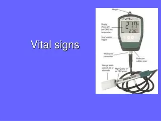
- Thermometers – 3 types • Glass mercury – mercury expands or contracts in response to heat. (just recently non mercury) • Electronic – heat sensitive probe, (reads in seconds) there is a probe for oral/axillary use (red) & a probe for rectal use (blue). There are disposable plastic cover for each use. Relies on battery power – return to charging unit after use. • Infrared Tympanic (Ear) – sensor probe shaped like an otoscope in external opening of ear canal. Ear canal must be sealed & probe sensor aimed at tympanic membrane – ret’n to charging unit after use.
- Sites (P&P p. 216)
- Assessing Radial Pulse • Left ventricle contracts causing a wave of bld to surge through arteries – called a pulse. Felt by palpating artery lightly against underlying bone or muscle. • Carotid, brachial, radial, femoral, popliteal, posterior tibial, dorsalis pedis P&P p. 226 • Assess: rate, rhythm, strength – can assess by using palpation & auscultation. • Pulse deficit – the difference between the radial pulse and the apical pulse – indicates a decrease in peripheral perfusion from some heart conditions ie. Atrial fibrillation.
- Procedure for Assessing Pulses • Peripheral – place 2nd, 3rd & 4th fingers lightly on skin where an artery passes over an underlying bone. Do not use your thumb (feel pulsations of your own radial artery). Count 30 seconds X 2, if irregular – count radial for 1 min. and then apically for full minute. • Apical – beat of the heart at it’s apex or PMI (point of maximum impulse) – 5th intercostal space, midclavicular line, just below lt. nipple – listen for a full minute “Lub-Dub” • Lub – close of atrioventricular (AV) values – tricuspid & mitral valves • Dub – close of semilunar valves – aortic & pulmonic valves
- Assess: rate, rhythm, strength & tension • Rate – N – 60-100, average 80 bpm • Tachycardia – greater than 100 bpm • Bradycardia – less than 60 bpm • Rhythm – the pattern of the beats (regular or irregular) • Strength or size – or amplitude, the volume of bld pushed against the wall of an artery during the ventricular contraction • weak or thready (lacks fullness) • Full, bounding (volume higher than normal) • Imperceptible (cannot be felt or heard) 0----------------- 1+ -----------------2+--------------- 3+ ----------------4+ Absent Weak NORMAL Full Bounding
- Normal Heart Rate
- Assess (cont.) • Tension – or elasticity, the compressibility of the arterial wall, is pulse obliterated by slight pressure (low tension or soft) • Stethoscope • Diaphragm – high pitched sounds, bowel, lung & heart sounds – tight seal • Bell – low pitched sounds, heart & vascular sounds, apply bell lightly (hint think of Bell with the “L” for Low)
- Respirations • Assess by observing rate, rhythm & depth • Inspiration – inhalation (breathing in) • Expiration – exhalation (breathing out) • I&E is automatic & controlled by the medulla oblongata (respiratory center of brain) • Normal breathing is active & passive • Women breathe thoracically, while men & young children breathe diaphramatically ***usually • Asses after taking pulse, while still holding hand, so pt is unaware you are counting respiratons
- Assessing Respiration
- Blood Pressure • Force exerted by the bld against vessel walls. Pressure of bld within the arteries of the body – lt. ventricle contracts – bld is forced out into the aorta to the lg arteries, smaller arteries & capillaries • Systolic- force exerted against the arterial wall as lt. ventricle contracts & pumps bld into the aorta – max. pressure exerted on vessel wall. • Diastolic – arterial pressure during ventricular relaxation, when the heart is filling, minimum pressure in arteries. • Factors affecting B/P • lower during sleep • Lower with bld loss • Position changes B/P • Anything causing vessels to dilate or constrict - medications
- B/P (cont.) P&P p. 240 see table 9-3 • Measured in mmHg – millimeters of mercury • Normal range • syst 110-140 dias 60-90 • Hypertensive - >160, >90 • Hypotensive
- B/P (cont.) • Cuff – inflatable rubber bladder, tube connects to the manometer, another to the bulb, important to have correct cuff size (judge by circumference of the arm not age) • Support arm at heart level, palm turned upward - above heart causes false low reading • Cuff too wide – false low reading • Cuff too narrow – false high reading • Cuff too loose – false high reading • Listen for Korotkoff sounds – series of sounds created as bld flows through an artery after it has been occluded with a cuff then cuff pressure is gradually released. P&P p. 240. • Do not take B/P in • Arm with cast • Arm with arteriovenous (AV) fistula • Arm on the side of a mastectomy i.e. rt mastectomy, rt arm
- Procedure – B/P
- Procedure (cont.)
- B/P Lower Extremity • Best position prone – if not – supine with knee slightly flexed, locate popliteal artery (back of knee). • Large cuff 1 inch above artery, same procedure as arm. Systolic pressure in legs maybe 10-40 mm hg higher • If unable to palpate a pulse – you may use a doppler stethoscope
- Oxygen Saturation (Pulse Oximetry) • Non-invasive measurement of oxygen saturation • Calculates SpO2 (pulse oxygen saturation) reliable estimate of arterial oxygen saturation • Probes – finger, ear, nose, toe • Patient with PVD or Raynauds syndrome – difficult to obtain. • Normal – 90-100% • Remove nail polish • Wait until oximeter readout reaches constant value & pulse display reaches full strength • During continuous pulse oximetry monitoring – inspect skin under the probe routinely for skin integrity – rotate probe.
- Procedure – Vital Signs
- Vital Signs (cont.)

Vital Signs
Vital Signs. Shurouq Qadose 17/2/2008. Vital signs are temperature, pulse, respiration, blood pressure and pain. A change in vital signs may indicate a change in health.
2.74k views • 74 slides

Vital Signs
Vital Signs. RTEC 93 Venipuncture for Radiographers. When do you wash your hands?. When hands are visibly soiled Before and after patient contact After removal of gloves After using the toilet After blowing or wiping the nose Upon leaving an isolation area How long do you wash? .
1.25k views • 29 slides
Vital Signs
Vital Signs. Vital Signs. Provide information about body function Include: temperature pulse respiration blood pressure Changes may be the first sign of disease Accuracy is imperative !. Vital Signs Temperature. Measurement of the balance between heat lost and heat produced
1.51k views • 42 slides
VITAL SIGNS
2. Vital Signs. Vital signs are an outward clue to what is going on in the patient's body. 3. Vital Signs. Baseline Vital Signs provide a basis for comparison of later sets of Vital Signs. 4. Vital Signs. It is important to monitor
780 views • 22 slides

Vital Signs
Vital Signs. Jarvis, Chapter 9. Vital Signs. Classic Vital Signs – TPR/BP Temperature Pulse Respirations Blood Pressure Additional Vital Signs Height Weight BMI (Kg/m2) or (702Xlbs/in2) Supine, orthostatic BP. Temperature. Measurement of metabolic activity Core vs Surface Exercise
949 views • 29 slides

Vital signs
2. Definition: There are four objective assessment data that indicate how well body is functioning and very sensitive to alteration in physiology. Body Temperature Pulse Respirations Blood Pressure . 3. Times to assess vital sign:. 1) On admission to health care agency to obtain base line data.2)
794 views • 46 slides

VITAL SIGNS
VITAL SIGNS. Professor Blakey NUR302. Vital Signs. Temperature Pulse Respirations Blood Pressure Health Status Changes Accuracy, Responsibility. Vital Signs. When are they reported? When are they recorded?. Temperature. Sites: Oral- Taken routinely Taken per MD order
1.24k views • 36 slides

VITAL SIGNS
VITAL SIGNS. Medical Foundations. Vital Signs (Signs of Life). Temperature Pulse Respirations Oxygen Concentration Pupils Blood Pressure. TEMPERATURE. afebrile a = without, febrile = fever hypothermia Below 95 0 F pyrexia Above normal temperature pyrogenic
571 views • 17 slides

Vital Signs
Vital Signs. Respirations. Be Professional. Breathing Patterns. Abnormal Breathing. Cheyne -Stokes respirations.
941 views • 15 slides

Vital Signs
Vital Signs. Respirations. Respirations. Process of taking in oxygen & expelling carbon dioxide 1 respiration = inspiration & expiration Measure while taking pulse; pt is not to know you are measuring resp. Leave fingers @ pulse site while counting respirations. Range of Respirations.
402 views • 7 slides

Vital Signs
Configuring RPMS-EHR for Meaningful Use R esource P atient M anagement S ystem. Vital Signs. Presenters: Angela Flanagan, BSN, RN, CDE OIT EHR Nurse Consultant Theresa Tsosie-Robledo, MSN, RN, BC OIT EHR Nurse Consultant and Informaticist. Core MU Objective.
335 views • 13 slides

Vital Signs
Vital Signs. Pulse Oximetry. Bellringer. Think back to the last time you or a family member went to see a doctor. What vital signs ( temperature, oxygen level, blood pressure, pulse rate, respirations ) did they take? How were these obtained? (with a machine or nurse take manually)
521 views • 9 slides

Vital Signs
Vital Signs. =Temperature =Pulse =Respiration =Blood Pressure =The fifth vital sign- observing and reporting the level of pain. Do not take an oral temperature on a person who…. Is unconscious Is using O 2 Is confused or disoriented Is paralyzed from stroke Has facial trauma
505 views • 12 slides
Vital Signs
Vital Signs. Created by Debbie Johnson RN- 2003. Vital Signs (VS). Temperature ( T) Pulse (P) Respiration (R) Blood Pressure (BP). Normal Range Temperature. Ideal Normal Range Oral 98.6 97.6-99.6 Axillary 97.6 96.6-98.6 Rectal 99.6 98.6-100.6 Tympanic 98.6 98.6.
673 views • 20 slides
Vital Signs
Vital Signs. Medical Science 1. Lesson Objectives. Understand What vitals are and how to document them Learn How to: Take Pulse Rate Take Respiration Rate Take Blood Pressure. What are vital signs?. Outward signs of what is going on inside of the body Pulse Respirations Blood Pressure
707 views • 18 slides
Vital Signs
Vital Signs. Guidelines for Measuring Vital Signs. Establish a baseline for future assessments. Be able to understand and interpret values. Appropriately delegate measurement. Communicate findings. Ensure equipment is in working order. Accurately document findings. Circulatory Needs.
1.26k views • 78 slides
Vital signs
Vital signs. Outline. Vital Signs Definition Temperature Pulse Rate Respiratory Rate Blood Pressure Pain. Vital sign. physical signs that provide data to determine a person’s state of health
633 views • 38 slides

Vital Signs
Vital Signs. Start. TABLE OF CONTENTS. Spirometer. Electrocardiogram. Click on a Topic. Vital Signs. Spirometer. Vital Signs. Electrocardiogram (EKG). EKG Phases: Ventricular Phases. EKG Phases: Comparison of Atrial and Ventricular Phases. Lead 2 EKG Components. Vital Signs.
435 views • 15 slides
